Vitamin A is actually a family of similar compounds, the retinoids, that are related to retinol. Apparently, retinoic acid performs most of the functions of vitamin A, binding to DNA receptors in target cells. You can see the diagram of this and it might make more sense.
There are 4 biologically important retinoids:
Retinol– the alcohol form. Retinol esters are packaged into chylomicrons and transported to the liver for storage. When retinol is released from the liver, it complexes with retinol binding protein (RBP) and is delivered to target tissues throughout the body (except heart and skeletal muscle). In the cytosol, retinol is irreversibly oxidized to retinoic acid (similar to steroid hormones) which binds to nuclear receptors forming a complex which activates gene transcription of protein products .
Retinal– the oxidized form of retinol. The two are readily convertable. Retinal is a component of the visual pigments within rod and cone cells of the retina (see below).
Retinoic Acid– the oxidized form of retinal. It cannot be reduced in the body. Thus, it cannot give rise to retinol or retinal. It has wide pharmacologic therapies in dermatology.
b-carotene– plant form of vitamin A. Oxidatively cleaved (via thyroid hormone) in the intestine to 2 molecules of retinal. Unfortunately, this conversion process is inefficient and the vitamin A activity of b-carotene is only about 1/6 that of retinol.
RDA: for adults- 1000 retinol equivalents (RE) for males and 800 RE for females. One RE=1 mg of retinol; 6 mg of b-carotene; or 12 mg of other carotenoids.
Good sources of Vitamin A: liver, kidney, cream, butter, and egg yolks have pre-formed vitamin A. Yellow and dark green vegetables and fruits are good sources of carotenes.
Functions of Vitamin A: The functions of vitamin A can be divided into four categories.
Visual Cycle- see this in the diagram below.
Growth- Absence of vitamin A prevents bone growth from keeping up with the growth of nervous tissue and can lead to CNS damage. Also, there is a hyper-keratinization of the taste buds on the tongue which decreases the person’s appetite (at first anyway).
Reproduction- prevents fetal resorption in the female and supports spermatogenesis in the male. Retinoic acid doesn’t help here or with the visual cycle. Therefore, if anyone has only had retinoic acid since birth, they are blind and sterile.
Maintenance of Epithelial Cells- you gotta have this stuff for epithelial differentiation and mucous secretion.
active forms of vitamin A are used in the treatment of skin disorders and acute progranulocytic leukemia (M3)
• topical tretinoin (all-trans-retinoic acid) is used in the treatment of psoriasis and mild acne
• oral isotretinoin is used to treat severe acne, however, it is teratogenic, so women must have a pregnancy test before it is prescribed and must be placed on birth control pills for the full duration of therapy 0 is used to treat acute progranulocytic (promyelocytic) leu emia (M3) and is thought to induce maturation of the leukemic cells
Retinoic acid as a pharmacotherapeutic- effective treatment of acne and psoriasis. Everything I have seen states that you have to use an all trans retinoic acid (tretinoin) which is too toxic for systemic use and must only be applied topically. However, if the pt doesn’t respond to this therapy, and if they have a severe case, you can use isotretinoin (13-cis retinoic acid) orally.
b-carotene- Populations that consume increased amounts of b-carotene also have a reduction in heart disease and lung and skin cancer. These effects are independent of b-carotene being a precursor to vitamin A and probably due to its antioxidant effects (see oxidative damage). b-carotene, unlike vitamin A, is not toxic even at high doses.
Toxicity: Some would argue that this can be the most toxic of the vitamins. You shouldn’t exceed 7.5 m. An excess of b-carotenes in the diet turns the skin yellow, however, the sclera remain white and signs of vitamin A toxicity do not occur
Deficiency Symptoms: Night blindness (nyctalopia), xerophthalmia (dryness of the cornea & conjunctiva – can lead to blindness), dry-rough skin, follicular keratosis, bronchogenic carcinoma (squamous metaplasia may progress to dysplasia & cancer), poor wound healing, & impaired bone growth. Vitamin A deficiency is the most common deficiency in the world and the 2nd leading cause of blindness (second only to Chlamydia trachomatis).
The active form of thiamine is thiamine pyrophosphate (TPP) which is essential for the conversion of pyruvate to acetyl CoA and Step 4 of the Kreb’s cycle. It is also essential for the conversion of glyceraldehyde-3-phosphate (one of the glycolysis intermediates) to ribose-3-phosphate.
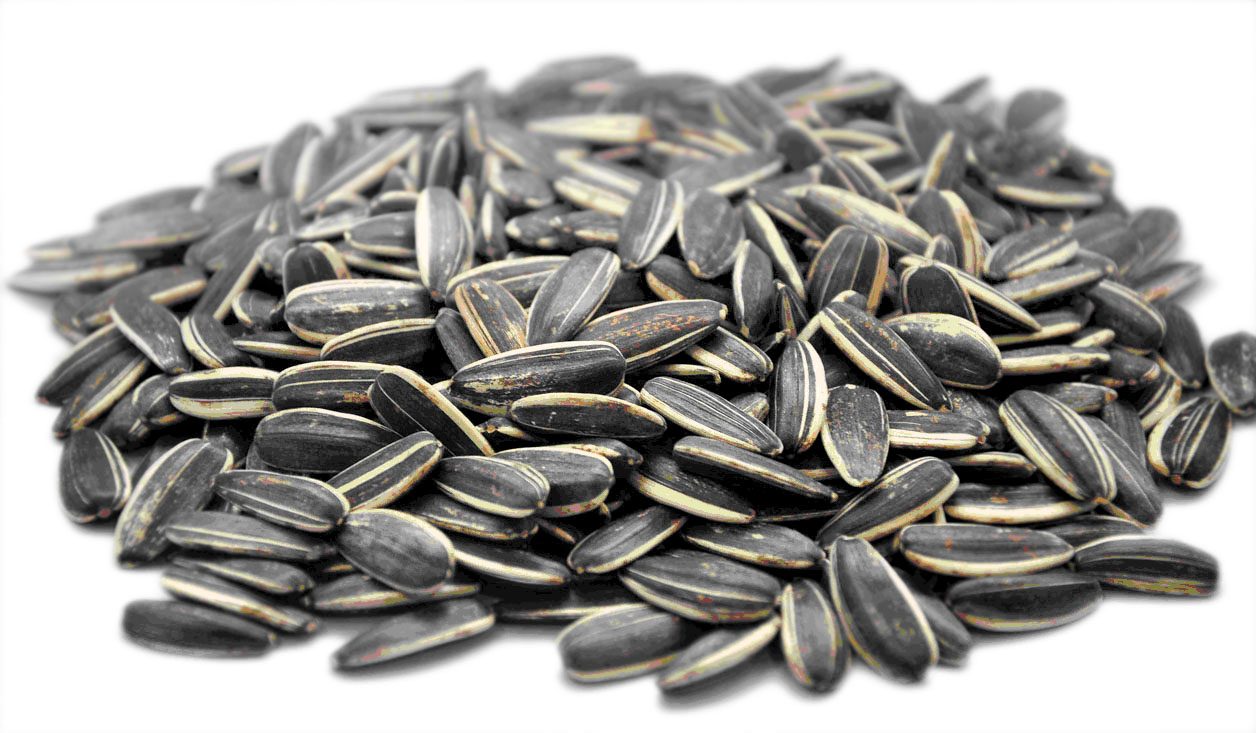
Good sources: Sunflower seeds, liver, pork, whole and enriched grains, and dried beans. According to one source, the outer layer of seeds is high in thiamine. Thus, whole wheat breads are high in thiamine while bread made from milled grain is low in thiamine.
DRI: 0.9-1.2 mg per day
Functions: the functions of this vitamin are listed above. It also assists in nerve function.
Cofactor to over 24 enzymes
Essential to the production of energy from food in the form of ATP (adenosine triphosphate)
Cofactor to pyruvate dehydrogenase (for energy production in Krebs cycle)
Cofactor in the maintenance of myelin sheath (nerve cell insulator)
Activates membrane ion channels which affect nerve and muscle cell function
Cofactor to transketolase for fat, protein and carbohydrate metabolism
Cofactor to transketolase for synthesis of ribose for incorporation into DNA (deoxyribonucleic acid)and RNA
Cofactor to transketolase for synthesizing cellular NADPH (Nicotinamide Adenine DinucleotidePhosphate Hydrogen) used for fatty acid biosynthesis
Cofactor to 2-oxo-glucarate dehydrogenase (also called α-ketoglutarate dehydrogenase) whichmakes acetylcholine, gamma amino butyric acid (GABA) and glutamate
Cofactor to α-ketoglutarate dehydrogenase for mitochondrial energy production
Cofactor to branched chain ketoacid dehydrogenase for mitochondrial energy production
Cofactor in the hexose monophosphate shunt, an anabolic pathway used by certain cells
Cofactor to glucose metabolism, particularly in central nervous system
Cofactor in the mitochondrial respiratory chain
Deficiency impairs oxidative metabolism leading to increased pyruvate and lactate, which cancause vasodilation, retention of water and contribute to heart failure (“cardiac or wet beriberi”)
Some cardiac medication decreases vitamin B1 uptake in myocytes (muscle cell)
Deficiency in children linked to delayed language development
Supplementation may benefit autistics
Supplementation may benefit inborn errors of metabolism of branched chain amino acids such as thiamin responsive branched chain ketoaciduria (also known as maple syrup urine disease)
Regulates intracellular p53 activity (a protein that protects against tumors)
Deficiency may cause serotonin depletion which can decrease pain threshold, decrease musclerepair enzymes (muscle soreness) and poor energy production (fatigue)
Key role in restorative sleep
Plays a role in acetylcholine production in the brain
Plays a role in nerve impulse conduction
Conversion of thiamin to biologically active form requires magnesium
Sulfites in food destroy vitamin B1
Excess alcohol inhibits activation of thiamine coenzymes
Deficiencies:Beri-Beri which means “I can’t, I can’t.” Infantile beri-beri symptoms include: tachycardia, vomiting, convulsions, and death. Adult beri-beri symptoms are: dry-skin, irritability, disorderly thinking, and progressive paralysis. Alcoholics are particularly at risk for thiamine deficiency for two reasons. First of all, they are likely to have a reduced intake and, secondly, alcohol decreases thiamine absorption.
Toxicity: Thiamine is not considered toxic. However, some have linked headaches and insomnia to thiamine when their doses exceeded 5 mg/d for 4-5 wks.
Riboflavin is needed for the formation of flavin mononucleotide (FMN, seen in Complex I of the ETS) and flavin adenine dinucleotide (FAD, from Step 6 of the Krebs). Below is a diagram that shows their structures.
Riboflavin is light-sensitive! That’s why you won’t see milk in a clear bottle or container.
Good sources: Milk, eggs, liver, green leafy veggies, and whole & enriched grains.
DRI: 0.9-1.3 mg per day
Functions: The 2 co-enzymes discussed above (FMN and FAD).
All known functions:
Essential to the production of energy from food in the form of ATP (adenosine triphosphate)
Active form is FAD (flavin adenine dinucleotide) and FMH (flavin mononucleotide, also calledriboflavin 5’-phosphate) which are cofactors for oxidation-reduction reactions in energy production
FAD assists methylation reactions throughout body
Coenzymes derived from riboflavin are called flavins
Enzymes that use a flavin coenzymes are called flavoproteins
Flavins metabolize drugs and toxins
Low B2 can impair methylation reactions in the brain, which may present clinically as depression
Activates glutathione reductase, which regenerates the antioxidant glutathione
Key role in iron utilization
Aids in mobilization of ferritin from tissues; helpful for anemia
FAD is a cofactor for methylenetetrahydrofolate reductase (MTHFR) so for people who arehomozygous for 677C→T MTHFR gene, riboflavin may lower their homocysteine
Recycles folate into a usable methyl-donor form (converts 5,10-methylene TH4-folate to 5-methylTH4-folate)
People with 677C→T MTHFR gene tend to respond well to B2 therapy to lower blood pressure
Cofactor to xanthine oxidase, which synthesizes uric acid and aids in purine catabolism
Cofactor to pyridoxal 5’-phosphate oxidase, which converts vitamin B6 into its active form
Cofactor in the conversion of retinol (vitamin A) to retinoic acid
Cofactor to kynurenine mono-oxygenase, which converts tryptophan into niacin containing enzymesNAD and NADP
Cofactor to NAD(P):quinine oxidoreductase which aids in detoxification and chemoprevention
Cofactor to protoporphyrinogen oxidase which synthesizes hemoglobin
Cofactor to dihydrolipoyl dehydrogenase which aids energy metabolism
Cofactor to fatty acyl-CoA-dehygrogenase which aids in fatty acid oxidation
Cofactor to succinate dehydrogenase which is used in the Krebs cycle for energy production
Cofactor to NADH dehydrogenase (also called ubiquinone oxidoreductase) which functions inmitochondrial respiration
Cofactor to sphinganine oxidase which synthesizes sphingosine (component of nerve tissue)
Cofactor to monoamine oxidase which aids in metabolism of several neurotransmitters such asserotonin, melatonin, epinephrine and norepinephrine
Role in mitochondrial energy metabolism may explain its efficacy in migraine prophylaxis
Role in glutathione reductase implicates riboflavin deficiency in cataract formation
Minimizes pain associated with inflammation
Augments the antinociceptive (painkiller) effects of morphine
Good sources: meats, liver, nuts, whole and enriched grains. Some niacin can be synthesized from tryptophan (inefficient).
DRI: 12-16 mg/d
Functions: Co-enzyme formation (NAD+ and NADP), fatty acid metabolism, and decrease serum cholesterol. It is the most potent agent to increase serum HDL at doses 100 times the RDA (but many people can’t tolerate the side effects). It does this by strongly inhibiting lipolysis in adipose tissue, which is the largest producer of circulating FFA’s. Decreased FFA’s are needed for the formation of VLDLs. LDLs are produced from VLDLs. Thus, niacin inhibits the formation of LDLs (indirectly). It is especially useful for the treatment of type IIb hyperlipidemia (both VLDL and LDL levels are increased).
Also called nicotinic acid or niacinamide or nicotinamide (not related to nicotine found in tobacco)
Nicotinic acid & niacinamide have identical vitamin properties but are different pharmacologically
Around 200 enzymes require niacin derived NAD or NADP enzymes (also called coenzyme I and
coenzyme II) usually for redox reactions
Coenzyme for NAD (nicotinamide adenine dinucleotide) and NADP (nicotinamide adenine
dinucleotide phosphate) which is needed to metabolize food
Increases the rate of NAD synthesis
NADP regenerates glutathione
Cofactor in the mitochondrial respiratory chain
NAD catabolizes carbohydrates, proteins, fats and alcohol
Cofactor to mitochondrial aldehyde dehydrogenase which metabolizes alcohol
Cofactor to glucose-6-phosphate 1-dehydrogenase which metabolizes glucose in red blood cells
Cofactor to dihydropteradine reductase which is involved in dopamine and serotonin synthesis
NADP synthesizes fatty acids and cholesterol
Only nicotinic acid influences blood lipid levels
Increases HDL (high density lipoprotein)
Lowers triglycerides by inhibiting diacylglycerol acyltransferase-2 (enzyme that synthesizes
triglycerides in liver)
Cofactor to long-chain-3-hydroxyacyl-CoA dehydrogenase which metabolizes fat for energy
Down regulates vascular cell adhesion molecules (VCAM)
Inhibits the oxidation of LDL (low density lipoproteins)
Decreases the highly atherogenic Lp(a) by reducing its rate of synthesis in the liver
Shifts lipoproteins from small, dense (atherogenic) to large, buoyant (non atherogenic)
Inhibits vascular inflammation and improves endothelial function independent of effect on lipids
Supresses cytokine-mediated induction of nitric oxide synthase, thus decreasing inflammation
Cofactor in mono-ADP-ribosyltransferase, which plays a role in cell signaling
Cofactor in poly-ADP-ribose polymerase (PARP), which aid in DNA replication and repair
Cofactor in ADP-ribosyl cyclase, which initiates release of calcium ions from inside cell
Maintains proper methylation of PARPgenes that suppress tumor formation and growth
Influences cellular response to DNA damage (cancer prevention)
Extends lifespan of human cells in vitro
Slows telomere attrition rate by reducing reactive oxygen species in mitochondria
Mediates cell signaling pathways important to the prevention of cancer
Regulates p53 activity in cells (a protein that protects against tumors)
Protect pancreatic beta cells in type 1 diabetics although high doses may impair glucose tolerance
Precursor to glucose tolerance factor which facilitates insulin binding
Pharmacological doses may enhance calming effect of GABA (gamma aminobutyric acid) in brain
Pharmacological doses may exacerbate gout or cause hepatotoxicity (liver damage)
Inhibits the enzyme tryptophan pyrrolase which breaks down tryptophan in the liver
Increases conversion of tryptophan to serotonin
Increases REM (rapid eye movement) sleep
Improves quality and quantity of sleep via its role in serotonin synthesis
Dilates blood vessels (may alleviate migraine)
Severe deficiency can cause anxiety
Increases adiponectin (weight loss hormone secreted by fat cells)
Niacin-bound chromium may help reduce body weight
Deficiencies: Clinical signs of early niacinamide deficiency include anorexia, muscular fatigue, indigestion, depression, insomnia, headaches, glossitis and skin lesions. Severe deficiency may lead to pellagra, with dermatitis, dementia, diarrhea (the 3 D’s of pellagra), tremors and sore (black) tongue. Deficiencies of thiamin, riboflavin and pyridoxine commonly accompany (or can cause) niacinamide deficiency.
Toxicity: Histamine reaction at doses >35 mg. Facial flushing is one of the symptoms of this.
It means “It’s everywhere.” Apparently they named it this because it can be found in a number of sources.
Good sources: Liver, broccoli, and egg yolks.
DRI: 4-5 mg/d. No RDA has been set.
Functions: It is the precursor to Co-enzyme A (CoA) as in acetyl CoA or succinyl CoA (see glycolysis or Kreb’s cycle).
Component of coenzyme A and acyl carrier protein (ACP), which facilitates metabolism of two- carbon units (acetyl groups) in the Krebs cyle
Coenzyme A is a cofactor in over 70 enzymatic pathways
ACP is cofactor to fatty acid synthase, which is required for fatty acid elongation
Essential to the production of energy from food in the form of ATP (adenosine triphosphate) via itsrole in transfer of acetyl groups for the metabolism of carbohydrates, fats and proteins
Acetyl groups are involved in synthesis of cholesterol, steroid hormones, porphyrin (hemoglobin), phospholipids (cell membranes)
Coenzyme A is involved in coenzyme Q10 synthesis
Coenzyme A is needed for prostaglandin synthesis
Coenzyme A is needed for acetylcholine synthesis
Coenzyme A is needed for melatonin synthesis
Coenzyme A is needed for pyruvate degradation
Coenzyme A is required for phase II detoxification of xenobiotics and several drugs by the liver
ACP is needed for sphingolipid production, makes the myelin sheath around nerves
Modulates protein structure, gene expression and cell signaling via coenzyme A activity
Increases activity of lipoprotein lipase, an enzyme that breaks down fat cells
May reduce hunger during calorie restriction
Modifies action of several enzymes involved in cholesterol synthesis
Favorably alters LDL (low density lipoprotein) metabolism to less atherogenic type
Functions: Biotin is required for proper metabolism of fats and carbohydrates. Biotin-dependent enzymes catalyze the addition of carboxyl groups (COO-) from bicarbonate, for use in fatty acid biosynthesis, gluconeogenesis, lipogenesis, propionate metabolism and leucine catabolism. Biotin is a co-enzyme in glucose production and fat synthesis.
Cofactor Functions:
Cofactor to acetyl-CoA carboxylase, needed for fatty acid metabolism
Cofactor to pyruvate carboxylase, an enzyme needed for gluconeogenesis
Cofactor to methylcrotonyl-CoA carboxylase, which metabolizes leucine
Cofactor to propionyl-CoA carboxylase (also called holocarboxylase synthetase), which metabolizes amino acids and cholesterol
Cofactor for fatty acid synthesis (specifically for elongating the fatty acid chain)
All Other Functions:
The protein in egg whites (avidin) binds to biotin preventing its absorption so those who consume raw egg are at increased risk of biotin deficiency
Essential cofactor for four carboxylase enzymes in the mitochondria
Common indications of biotin deficiency include brittle nails, alopecia and dermatitis due to impaired fatty acid synthesis from reduced biotin-dependent carboxylases enzymes (particularly acetyl-CoA carboxylase)
Decreases synthesis of cytokines (interleukin-1 b and interleukin-2)
Decreases proliferation of peripheral blood mononuclear cells
Affects gene transcription (over 2,000 biotin dependent genes have been identifies)
Biotin deficiency in pregnancy may be teratogenic
Stimulates glucose-induced insulin secretion in pancreatic beta cells
Induces glucokinase, an enzyme needed for cells to use glucose
Accelerates glycolysis in liver and pancreas
Studies indicate in can reduce triglycerides and improve glycemic control
Biotin deficiency has induced depression in animal and human studies
Animal studies show induced biotin deficiency presents clinically as fatigue
Reduces blood pressure by activating the enzyme guanylate cyclase which in turn activates cGMP (cyclic guanosine monophosphate)
Anticonvulsant therapy such as phenobarbital and valproic acid may induce biotin deficiency
Deficiencies: Symptoms of biotin deficiency include erythematous exfoliative dermatitis, thinning hair, fatigue, irritability, mild depression, somnolence, muscle pains, anorexia, nausea, mild anemia. Infants with seborrheic dermatitis, Leiner’s disease or alopecia may indicate a biotin deficiency, along with symptoms of ketoacidosis, poor feeding, vomiting, lethargy, coma and developmental retardation. Dietary symptoms include fatigue, dry skin, body hair loss, nausea, loss of appetite and mild depression.
Those at risk for biotin deficiency include: persons consuming excessive amounts of raw egg whites, inherited disorders of biotin metabolism, extended total parenteral nutrition (biotin-free), loss of enteric gut microflora from antibiotic
therapy or altered gut motility, pregnant and lactating women, antiepileptic drug therapy, alcoholics, trauma (burns and surgery), elderly, malabsorption (especially achlorhydria).
Vitamin B6 is actually composed of a group of chemicals: pyridoxine, pyridoxal, and pyridoxamine. Pyridoxine is found in plants and the other two are found mainly in animals. Pyridoxal phosphate is the biologically active co-enzyme.
DRI: 1-1.7 mg/d. However, the requirement for pyridoxine goes up when you have a high protein intake.
FUNCTIONS: Vitamin B6 is needed to metabolize proteins and is important for a healthy immune system, nerves, bones and arteries. Vitamin B6 is a complex of three similar molecules: Pyridoxine, Pyridoxal and Pyridoxamine. All are present in foods and converted into pyridoxal-5-phosphate, the most active coenzyme form. The primary functions of vitamin B6 are in protein metabolism, transferring amino acid and sulfur groups. Roles in synthesis of heme (for hemoglobin), niacin, neurotransmitters, connective tissues, eicosanoids and sphingolipids in nerve sheaths are also essential. Vitamin B6 also participates in the utilization of glycogen and immune function.
Cofactor Functions:
Cofactor to 112 known enzymes
Cofactor for dopa decarboxylase which converts L-dopa to dopamine and 5HTP to serotonin
Cofactor to alanine-glyoxalate aminotransferase which converts glyoxylate to glycine
Cofactor to erythrocyte alanine aminotransferase which transfers amino groups
Cofactor to aspartate aminotransferase which moves amino groups between aspartate & glutamate
Cofactor to glycogen phosphorylase which releases glucose from glycogen
Cofactor to ornithine aminotransferase which makes proline & prevents gyrate atrophy (retinal degeneration)
Cofactor to glutamic acid decarboxylase that converts glutamate to the neurotransmitter GABA
Cofactor in the utilization of selenium (disconnects selenium from selenoproteins for use in the body)
Conversion to PLP is vitamin B2 dependent; Deficiency of B2 impacts B6 function
Cofactor in the synthesis and function of several neurotransmitters including serotonin, gamma-amino-butyric acid (GABA), dopamine, epinephrine and norepinephrine
Cofactor in mitochondrial respiratory chain to produce energy via ATP (adenosine triphosphate)
Cofactor to enzymes that converts homocysteine to cysteine (cystathionine synthase and cystathionase)
Cofactor in the synthesis of taurine
Cofactor in the synthesis of heme (hemoglobin)
Cofactor in the metabolism of vitamin B3 (niacin) from tryptophan via kynurenine pathway
Cofactor in the synthesis of connective tissue and eicosanoids
Cofactor in the synthesis of sphingolipids for nerve cell insulation
Cofactor in the synthesis of antibodies (key role in immune function)
Cofactor for tyrosine decarboxylase, which catalyzes the conversion of tyrosine to tyramine
Cofactor to lysyl oxidase which builds arterial integrity via role in collagen and elastin structure
Cofactor to aminolevulinic acid synthase which aids in hemoglobin synthesis
Cofactor to serine hydroxymethyltransferase which transfers methyl groups from serine to folate and initiates immune cell proliferation
Cofactor to serine palmitoyltransferase which makes sphingolipids for nerve cell insulation
Cofactor to serine facemase which synthesized neurotransmitter D-serine
Cofactor to sphinosine-1-phosphate lysase which makes sphingolipids for nerve cell insulation
Cofactor to cystathionine-b-synthase which metabolizes homocysteine and serine to form cystathionine
Cofactor or kynureninanse which metabolizes tryptophan into vitamin B3 for NAD cofactors
Cofactor to GABA aminotransferase which breaks down GABA
Cofactor to diamine oxidase, which catabolizes exogenous histidine in the gut
Cofactor for histidine decarboxylase which converts histidine to histamine
All Other Functions:
B6 is a complex of three molecules: pyridozine, pyridoxal, and pyridoxamine which are all converted to pyridoxal-5-phosphate (PLP), the active coenzyme form
Bins with histamine and inactivates it (may mitigate allergic response in asthma)
Increases peripheral metabolism of levodopa (l-dopa) making it less available for uptake into the brain (may diminish effectiveness of l-dopa medication for Parkinson’s patients when not given with carbidopa which is a peripheral decarboxylase inhibitor)
High dose B6 can be as effective as Ritalin for ADHD due to its effect of increasing serotonin
Synergistic effect with magnesium for autism patients
Increases intracellular uptake of magnesium and vice versa
Low B6 linked to high CRP (C-reactive protein), a marker of inflammation
Supplementation suppresses pro-inflammatory cytokines such as IL-6 and TNF-a
Tissue specific depletion of B6 occurs during inflammation
Crucial for DNA methylation thus regulating gene expression
Deficiency impairs conversion of alpha-linolenic acid to EPA and DHA
Protects genes from estrogen-induced damage (detoxifies estrogen) lowering risk of hormone related cancers
Regulates sex hormones and binds to steroid hormone receptors, thus decreasing their effects
Reduces prolactin levels which stimulates hypothalamus to increase testosterone
Inhibits pituitary (and other tissue) tumor proliferation via role in apoptosis (programmed cell death)
DEFICIENCIES: Convulsion, nausea, flaky skin, HA, insomnia (uncommon). Isoniazid can induce a deficiency, so you have to supplement with isoniazid prescription. Deficiency is rare but has been seen in infants with low intake of B6, females on OCP’s, and alcoholics.
Early vitamin B6 deficiency symptoms are primarily peripheral neuropathy, weakness, irritability, depression, insomnia and anxiety. More severe deficiency leads to dermatitis, nausea, vomiting and convulsions. Carpal tunnel syndrome,
premenstrual tension syndrome and atherosclerosis may also be related to vitamin B6 deficiency. Sideroblastic anemia is indicative of vitamin B6 deficiency. Homocysteine levels in serum may be elevated by a vitamin B6 deficiency.
TOXICITY: Nerve damage (it is the most toxic water soluble vitamin) @ doses > 200mg. Substantial improvement, but not complete recovery, occurs when the vitamin is removed. Another source indicated neurologic symptoms at doses of 2 grams/d.
GOOD SOURCES: Green leafy veggies, liver, lima beans, and whole grain cereals
DRI: 300-400 mg/d (0.3-0.4 mg/d); for women who are on birth control pills, this increases dramatically. After stopping BCPs, they must take 800 mg/d for at least 30 days before getting pregnant.
FUNCTIONS: Folic acid is essential for the biosynthesis of the purines and the pyrimidine , thymine.It is converted to tetrahydrofolic acid (THF) which is the biologically active form. THF receives one-carbon fragments from donors (ie. serine, glycine, & histidine) and transfers them to intermediates in the synthesis of amino acids and the nucleotides mentioned above. See the diagram below.
All Nutrient Functions:
High stomach pH (low stomach acid) decreases folic acid absorption
Needed for the production of red blood cells
Needed for the synthesis of nucleic acids
Key role in one carbon metabolism (methylation reactions) as it accepts & donates methyl groups
Precursor to SAMe (S-adenosylmethionine)
Influences telomere length via DNA methylation
Cofactor in the metabolism of methionine, histidine, tryptophan, glycine, serine, and formate
Required for the conversion of homocysteine into methionine
Affects noradrenaline and serotonin receptors in the brain
Inhibits NMDA receptors
Building block in synthesis of serotonin, dopamine, and norepinephrine
Low folate causes poor response to anti-depressant medications
Improves response to the anti-depressant drug fluoxetine
The lower the folate, the more severe the depression in some individuals
May improve response to erectile dysfunction meds (PDE5 inhibitors)
Cofactor in mitochondrial respiratory process
Improves endothelial function
Increases nitric oxide (NO) production in vascular endothelial cells
Cofactor to enzyme nitric oxide synthase
Enhances the availability of nitric oxide cofactors such a tetrahydrobiopterin
Improves flow mediated dilation
Lowers risk of colonic neoplasia in patients with ulcerative colitis
Rapidly dividing cells are the most vulnerable to folate deficiency
Alcohol interferes with the absorption and metabolism of folate
Methotrexate (cancer, arthritis, and psoriasis drug) is a folate antagonist
Individuals with homozygous C–> T in the C677T MTHFR (methylene tetrahydrofolate reductase) gene have higher folate requirements that those without two copies of this gene
Synthesis of active form of folic acid requires B2, B3, B6, zinc, Vitamin C and serine
Folate supplementation may increase Vitamin B12 requirements or aggravate neurological symptoms stemming from B12 deficiency
DEFICIENCIES: Folic acid deficiency is probably the most common vitamin deficiency in the U.S. (especially amoung pregnant females and alcoholics). Deficiency is manifested two ways: the first is Megaloblastic anemia because there isn’t enough synthesized nucleotides; the second is developmental abnormalities of the neural tube in the fetus.
The anemia is due to diminished synthesis of purines and thymidine. Therefore, the erythropoeitic cells are not able to make DNA or divide. Vitamin B12 deficiency can also cause this disorder (but at a different step). Make sure to evaluate the exact cause of megaloblastic anemia before instituting therapy. Deficiency can be brought on by increased demand (as in pregnant or lactating women), poor absorption, alcoholism, or medications (that inhibit dihydrofolate reductase such as methotrexate).
Neural tube development of the fetus is critically dependant on folic acid. Women considering getting pregnant should ensure intake of 0.4 mg/d. This amount must be consumed early because the neural tube develops very early in development (before most women know that they are pregnant). Supplementation should not exceed 1 mg/d to avoid complicating a vitamin B12 deficiency.
Deficiency Functions:
Deficiency causes neural tube defects such as spina bifida and cleft palate in newborns
Deficiency linked with cancers, especially colon and breast, although excess supplementation of folic acid has been linked with cancer cell proliferation and estrogen-related breast caner
Deficiency reduces estrogen and circulating testosterone
Deficiency alters cholinergic metabolism in the brain
Deficiency reduces proliferation of lymphocytes
TOXICITY: Masks vitamin B12 deficiency (mainly in elderly)
In contrast to the other water soluble vitamins, cobalamin is stored in the body (~4-5 mg).
There are 2 primary forms with which most people supplement: cyanocobalamin and methylcobalamin. Hydroxycobalamin is a stable form that is converted to the active, methylcobalamin form.
How it absorbs
When taken orally, Vitamin B12 binds to intrinsic factor which is produced in the stomach. The complex of Vitamin B12 & intrinsic factor is absorbable. Without intrinsic factor we cannot absorb Vitamin B12 through the gastrointestinal tract.
This issue is the most common reason that people don’t have adequate levels. We also commonly see deficiency in vegetarians and vegans because there are not good plant sources of B12.
Autoimmune attack of the gastric parietal cells destroys the bodies ability to produce intrinsic factor.
The B12-Intrinsic Factor complex is absorbed in the ileum of the small intestine. Some people may also have a gut disorder that could decrease the absorption of B12.
Good sources
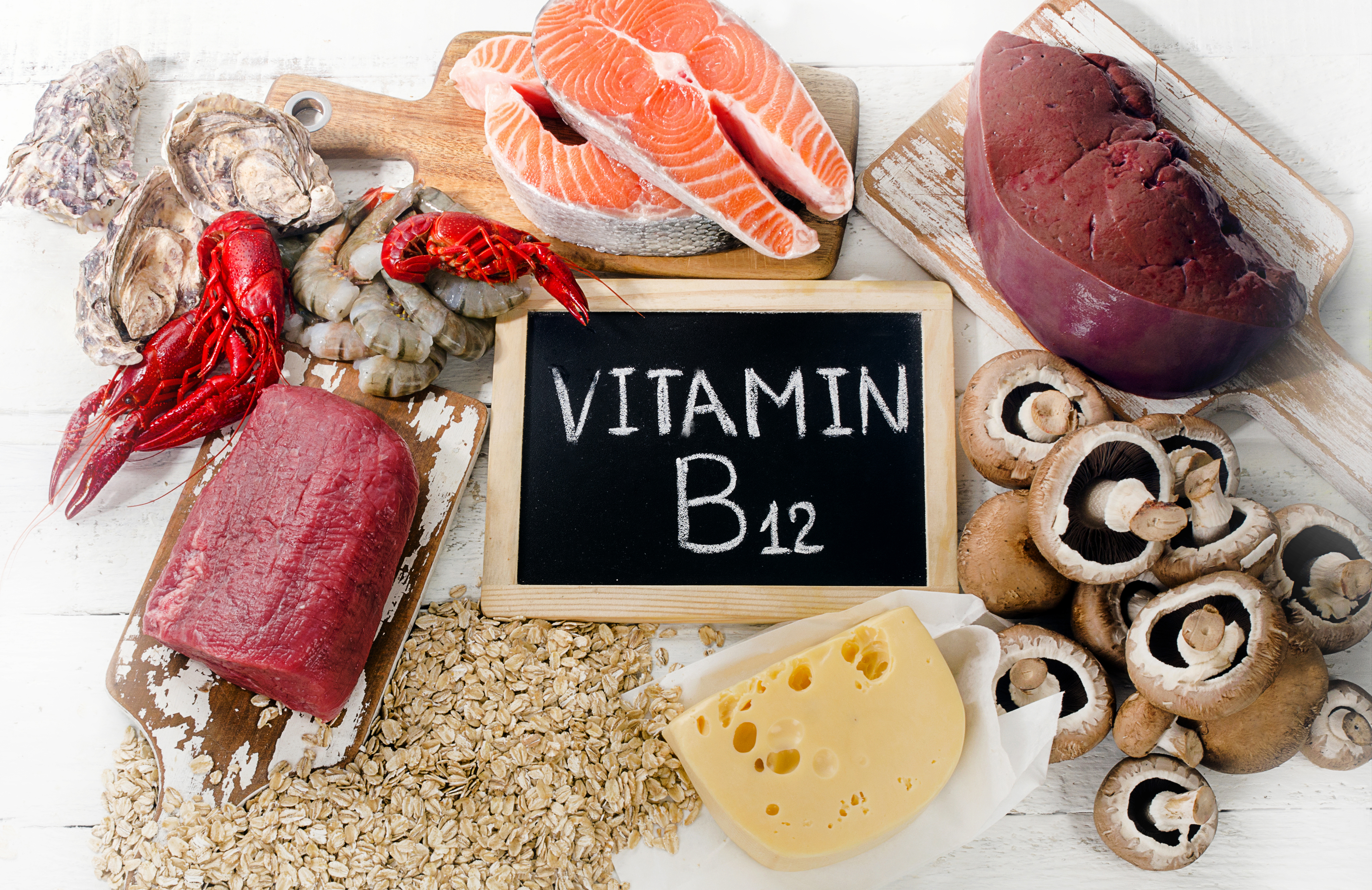
This vitamin is not found in plants! It is synthesized only by microorganisms. Animals obtain cobalamin from intestinal flora or by eating the meats of other animals. Especially liver, whole milk, eggs, oysters, fresh shrimp, pork, and chicken.
DRI: 1.8-2.4 mg/d
Our sublingual methylcobalamin is 2.5mg per day and also contains 5-MTHF 1mg. We also give methylcobalamin as an injection and it is included in several of our IV nutritional formulas.
Functions:
Vitamin B12 (cobalamin) is essential for 2 enzymatic reactions: synthesis of methionine and isomerization of methylmalonyl CoA that arises from odd fatty acid chains. Cobalamin converts some forms of TetraHydroFolate (THF) to the active form (5-MTHF). If cobalamin is deficient, the inactive forms build up and cannot be converted. Therefore, cobalamin deficiency can resemble folic acid deficiency and vice versa. This process is further discussed on the Methylation post.
Repletion improves treatment response in depression
Inhibits proliferations of malignant cells in vivo
May enhance efficacy of methotrexate
Protects retinal neurons against N-methyl-D-aspartate receptor mediated glutamate neurotoxicity
Deficiencies:
Pernicious anemia. Deficiency is most often due to an inability to absorb the vitamin rather than an inadequate amount in the diet. Vitamin B12 deficiency can also lead to CNS symptoms. Folic acid supplementation can correct the anemia associated with vitamin B12 deficiency but not the CNS effects. Make sure you know which is deficient!
Deficiency causes excess methylmalonic acid which decreases fatty acid synthesis for nerve cells.
Toxicity: None known
I often look at methylcobalamin as a ‘litmus test’ of the quality of nutritional supplements. It is slightly more expensive to produce so it helps me to understand how important quality may be to the manufacturer. I only recommend methylcobalamin, adenosylcobalamin, or hydroxycobalamin supplementation whether it be orally or injectable. Most traditional doctors use the cyanocobalamin version in their ‘B12 injections.’
Cyanocobalamin has a cyanide molecule attached to it. Do you want to inject your body with cyanide?
We recommend a supplement with B12 (methycobalamin) in a sublinqual form. Sublinqual forms are absorbed directly into the blood system so we don’t have to worry about intrinsic factor and gut health for absorption.
Vitamin B12 refers primarily to cyanocobalamin (they are used interchangeably), but also to several other cobalamins which possess similar nutritional properties. Vitamin B12 is synthesized by bacteria and is found in soil and in contaminated water. The average Western diet contains 5 to 15 μg/day of vitamin B12, and exceeds the recommended dietary allowance of 2 μg/day.1,2 The primary dietary sources of vitamin B12 are in foods of animal derivation (meat, eggs, and milk). Absorption of vitamin B12 occurs when it is bound to intrinsic factor from the stomach parietal cells.2 Vitamin B12 deficiency is rare in individuals who consume animal products unless there is an underlying vitamin B12 malabsorption condition. Strict vegetariansand babies of mothers who are strict vegetarians are at an increased risk for developing anemia.1-3 The body stores a large amount of vitamin B12 (i.e., 2-5 mg), primarily in the liver. It takes 2 to 5 years to develop a vitamin B12 deficiency even in the presence of severe malabsorption.1,2 However, it is estimated that 5% to 40% of the elderly population have B12 deficiency,4 probably due to reduced intrinsic factor production.2,5
Clinical Interpretation
Vitamin B12 deficiency, related to poor intestinal B12 absorption or dietary deficiency, is associated with pernicious anemia, cardiovascular disease, cancer, and neurodegenerative disorders.6 A cobalamin-dependent reaction involves the synthesis of methionine from homocysteine using methylcobalamin as a cofactor. One hypothesis is that this reaction has primary importance in vitamin B12 and folate deficiencies. A deficiency of either vitamin impairs tetrahydrofolate production.7 In megaloblastic anemia patients, this causes a defect in DNA synthesis that prevents cell division in the marrow. RNA synthesis and cytoplasmic component synthesis are not affected, which results in the production of large erythrocytes.1,2 In addition, a deficiency of either vitamin will result in homocysteine accumulation.1
A cobalamin deficiency may also cause increased methylmalonyl-CoA and its hydrolysis product, methylmalonic acid (MMA).1,8 MMA is regarded as a primary marker of cobalamin deficiency and there are increasing data that high homocysteine concentrations may contribute to occlusive vascular disease, cognitive dysfunction, and adverse pregnancy outcomes and malformations.7,9,10 Functional vitamin B12 deficiency in vegetarians may contribute to hyperhomocysteinemia in this population.3
Homocysteine and MMA are sensitive markers of cobalamin status and are used for the diagnosis and follow-up of cobalamin deficiency. Homocysteine is also elevated in folate deficiency and is used as an indicator of this deficiency state. MMA is a sensitive but specific marker of cobalamin function, although renal dysfunction may also cause significantly elevated MMA concentrations.9 Homocysteine is elevated in both folate and cobalamin deficiencies and also in pathologic states such as renal failure, thyroid dysfunction, coronary artery disease, and the acute phase after a cardiovascular event.7,9,10
A vitamin B12 test is used to check for megaloblastic anemia and to identify the cause of certain dementias or other nervous system symptoms (i.e., peripheral neuropathy). Vitamin B12 is usually measured at the same time as folic acid because a megaloblastic anemia may be caused by a deficiency of either vitamin. The two metabolic markers MMA and homocysteine are generally considered more sensitive indicators of vitamin B12 status than are plasma cobalamin levels.9,11 Homocysteine is an intermediary amino acid formed during the metabolism of methionine, an essential amino acid derived from protein. Hyperhomocysteinemia (> 15 µmol/L) induces vascular endothelial dysfunction and is considered to be an independent risk factor for atherosclerosis and cardiovascular disease (CVD). Treatment with folic acid, pyridoxine hydrochloride (vitamin B6), and vitamin B12 has been shown to reduce homocysteine levels in intervention studies and to reverse endothelial dysfunction independent of the effect of lowering homocysteine levels.11-14 Therefore, it is reasonable to propose that lowering homocysteine levels with folic acid and B vitamin supplements may help to decrease hyperhomocysteinemia.7,10-12
The most common screening test for vitamin B12 deficiency is the measurement of total plasma or serum
vitamin B12. Although cutoffs vary among assays and laboratories, a total vitamin B12 concentration < 148 pmol/L (< 200 pg/mL) is commonly considered indicative of vitamin B12 deficiency.1,2,5 High levels of vitamin B12 can occur in liver disease (such as cirrhosis or hepatitis) and some types of leukemia. However, the vitamin B12 test is not usually used to diagnose these conditions. Rarely, high levels may be found in obese or diabetes patients.
Causes of vitamin B12 deficiency include malabsorption, low dietary intake, strict vegetarian diet, pernicious anemia, gastric bypass or gastrectomy, ileal disease or resection, pancreatic insufficiency, absence or dysfunction of intrinsic factor, bacterial overgrowth, and impaired utilization.1,2
The most common symptoms of anemia include fatigue, shortness of breath, and palpitations. Vitamin B12 deficiency anemia may also be caused by a lack of intrinsic factor—pernicious anemia. The patient’s digestive system cannot absorb B12 properly. Signs and symptoms of pernicious anemia may include: a sore mouth and/or tongue, weight loss, pale or yellowish skin, diarrhea (sporadic), menstrual problems, greater susceptibility to infections.1,11 If the deficiency continues untreated the patient may have neurological signs and symptoms (e.g., tingling or numbness of the fingers or toes, general muscle weakness, difficulty walking, irritability, confusion, forgetfulness, tender calves).11
Who should be tested?
Pernicious anemia patients; approximately 60% have low RBC folate levels because vitamin B12 is required for normal transfer of MTHF from plasma to RBCs
Patients with elevated homocysteine
Patients post gastric bypass
Patients suspected of having macrocytic (megaloblastic) anemia; symptoms are weakness, fatigue, difficult concentration, irritability, headache, palpitations, and shortness of breath
Patients with peripheral arterial disease (PAD)
Patients with cognitive dysfunction, depression, mental changes, dementia, or insomnia
Patients with inadequate intake (e.g., malnutrition, malabsorption), increased demands (e.g., pregnancy, infancy), diseases associated with rapid cellular proliferation (hemolysis, leukemia, exfoliative dermatitis), jejunal diseases, short-bowel syndrome, and bacterial overgrowth
Patients on a strict vegetarian diet who may be deficient
Patients with chronic inflammatory bowel diseases
Patients with alcoholism, which may cause significant malnutrition and folate transport defects
Drugs (e.g., sulfasalazine, phenytoin, primidone, phenobarbital, oral contraceptives, methotrexate, triamterene) may reduce folic acid absorption and enhance folate deficiencies when patients on these drugs are not receiving supplementation
Treatment Considerations
Vitamin B12 and folate deficiencies usually require lifelong treatment with supplements. Lack of gastric intrinsic factor necessitates vitamin B12 injections. Once the diagnosis of vitamin B12 deficiency has been confirmed efficient treatment can be ensured either by injections every 2-3 months or by a daily dose of 1 mg vitamin B12.11 Patients who are taking metformin are at increased risk for vitamin B12 deficiency and may benefit from sublingual vitamin B12 supplementation.15 Vitamin B12 administration will increase red blood cell production, which may increase the need for iron supplementation as well.11
References
Snow CF: Laboratory diagnosis of vitamin B12 and folate deficiency; a guide for the primary care physician. Arch Intern Med 1999;159:1289-1298
Jordan NS: Hematology: Red and white blood cell tests. In: Traub SL, ed: Basic Skills in Interpreting Laboratory Data, 2nd ed. Bethesda, MD: American Society of Health System Pharmacists, 1996. pp. 302-304.
Herrmann W, Schorr H, Purschwitz K, et al. Total homocysteine, vitamin B12, and total antioxidant status in vegetarians. Clin Chem 2001;47(6):1094–1101.
Loikas S, Koskinen P, Irjala K, et al. Vitamin B12 deficiency in the aged: a population-based study. Age and Ageing 2007;36:177–183.
Miller JW. Assessing the association between vitamin B12 status and cognitive function in older adults. Am J Clin Nutr 2006;84:1259–60.
Ryan-Harshman M, Aldoori W. Vitamin B12 and health. Can Fam Physician 2008;54(4):536-41.
Stanger O, Herrmann W, Pietrzik K, et al. Clinical use and rational management of homocysteine, folic acid, and B vitamins in cardiovascular and thrombotic diseases. Z Kardiol 2004;93:439-453
Christen WG, Glynn RJ, Chew EY, et al. Folic acid, pyridoxine, and cyanocobalamin combination treatment and age-related macular degeneration in women. The Women’s Antioxidant and Folic Acid Cardiovascular Study. Arch Intern Med 2009;169(4):335-341.
Monsen AL, Ueland PM. Homocysteine and methylmalonic acid in diagnosis and risk assessment from infancy to adolescence. Am J Clin Nutr 2003;78:7-21.
Selhub J. The many facets of hyperhomocysteinemia: studies from the Framingham cohorts. J Nutr 2006;136:1726S-1730S.
Hvas AM, Nexo E. Diagnosis and treatment of vitamin B12 deficiency. An update. Haematologica 2006;91:1506-1512.
Homocysteine Lowering Trialists’ Collaboration. Dose-dependent effects of folic acid on blood concentrations of homocysteine: a metaanalysis of the randomized trials. Am J Clin Nutr 2005;82(4):806-812.
Moens AL, Vrints CJ, Claeys MJ et al. Mechanisms and potential therapeutic targets for folic acid in cardiovascular disease. Am J Physiol Heart Circ Physiol 2008;294:H1971-H1977.
Title LM, Cummings PM, Giddens K, et al. Effect of folic acid and antioxidant vitamins on endothelial dysfunction in patients with coronary artery disease. J Am Coll Cardiol 2000;36(3):758-65.
Moore EM, Mander AG, Ames A, et al. Increased risk of cognitive impairment in patients with diabetes is associated with metformin. Diabetes Care 2013;36(10):2981-7.
Vitamin C is a very important nutrient for a number of reasons. It is essential for several functions in the body which I’ve listed below. It is extremely safe. In fact, in the absence of kidney damage there are no known toxicities, especially if taken IV.
Good sources: Rapid growing veggies (cabbage, broccoli, asparagus, brussel sprouts), citrus fruits, and raw potatoes, organ meats (liver)
RDA: 60 mg/d. There is an active pool in the body of about 1500 mg. There is a limit to Vit C absorption. If you consume 100 mg then 100% is absorbed. However, if you consume 12,000 mg then only 16% is absorbed. However, IV Vitamin C can achieve very high serum levels without toxicity signs or symptoms.
Functions: Vitamin C is required for several metabolic functions in the body. One of its major roles is in the synthesis of collagen and elastin, the main structural proteins of skin, cartilage and blood vessels. It is also necessary in the production of several stress response hormones including adrenalin, noradrenalin, cortisol and histamine, and it is required in the synthesis of carnitine, an amino acid that facilitates the conversion of fatty acids into energy within the mitochondria. Vitamin C protects against heart disease in several ways: it helps dissolve arterial plaque, reduces free radical oxidation of cholesterol, decreases levels of the atherogenic lipoprotein Lp(a), and maintains the elasticity of vascular walls which helps control hypertension. In addition, vitamin C boosts immunity by increasing production of white blood cells, increasing levels of antibodies and interferon and modulating prostaglandin synthesis. It enhances iron absorption, promotes efficient wound healing, and detoxifies the body by binding to certain heavy metals so they can be eliminated from the body. The anti-cancer effects of vitamin C stem from its role as a potent, water-soluble antioxidant in the plasma and cytoplasm. It also protects nucleic acids (DNA) from oxidative damage and inhibits the formation of nitrosamines (carcinogenic compounds formed in the digestive tract). Additionally, it can regenerate vitamin E and works synergistically with other antioxidants such as beta-carotene and glutathione to increase their overall antioxidant effect.
Antioxidant (see oxidative damage) – The plasma ascorbate concentration in an oxidatively stressed patient (less than 45 µmol/L) measured is lower than healthy individual (61.4-80 µmol/L) according to McGregor and Biesalski (2006). Increasing plasma ascorbate level may have therapeutic effects in oxidative stress. Individuals with oxidative stress and healthy individuals have different pharmacokinetics of ascorbate
Vitamin C donates electrons in enzymatic reactions and quenches free radicals in plasma & cytoplasm
Replenishes the antioxidant Glutathione
This protects DNA from damage
Reduces free-radical oxidation of cholesterol
Involved in numerous catalyst reactions
Collagen & elastin synthesis
Vitamin C is a co-enzyme in hydroxylation reactions. If you will recall, proline and lysine are hydroxylated to form collagen.
Promotes wound healing
Increases absorption of non-heme iron
Enhances immune function
Increases production of white blood cells and interferon
Modulates prostaglandin synthesis
Decreases levels of creatine kinase after exercise
Improves blood vessel dilation
Catalyzes conversion of dopamine to norepinephrine
Necessary for the synthesis of epinephrine, norepinephrine, cortisol, & histamine
Large doses (>2 grams) have an antihistamine effect
Inhibits histamine-induced constriction of bronchial airways
Required in the synthesis of epinephrine
Regulates GABA (Gamma Amino Butyric Acid) receptors in the central nervous system (CNS)
Cofactor for carnitine synthesis
Cardiovascular protection
Metabolizes cholesterol to bile acid conversion
Dissolves arterial plaque
May decrease the atherogenic potential of Lp(a)
Lowers glycosylated hemoglobin (HbA1c), fasting and post-prandial glucose levels in diabetic patients
Required for the synthesis and metabolism of tyrosine
Aids in the detoxification of heavy metals
Inhibits the formation of nitrosamines (carcinogenic compounds) in the stomach
Partially restores thyroid function when liver detoxification ability is compromised
Deficiency alters methylation patterns in cancer cells
Enhances the effect of clomiphene, a fertility drug
Estrogen-containing contraceptives lower vitamin C levels
Increases estradiol (E2) in women on hormone replacement therapy
Lowers aromatase in the ovaries
Lowers serum uric acid levels
Catalyzes reactions that regulate oxytocin, vasopressin, and cholecystokinin
Anti-cancer effects
Slows age-related telomere shortening in human skin cells
Protects prostate against testosterone-induced tumors
Anti-cancer effects include formation of collagen to “wall off” tumors
Inhibits hyaluronidase, an enzyme that breaks down the matrix of cell walls
High Vitamin C intake can inhibit copper absorption
enhances chromium uptake
Regenerates Vitamin E. Supplementation with large amounts of either Vitamin C or Vitamin E increases the requirements of the other
Deficiencies: Deficiency symptoms include capillary fragility (due to its role in collagen formation) which often manifests clinically as bleeding/spongy gums, loose teeth, anemia, easy bruising, tender joints, muscle weakness and poor wound healing. Subclinical deficiency can also result in lowered immunity, anemia (due to vitamin C’s ability to enhance iron absorption) and fatigue (due to its role in the synthesis of carnitine and certain hormones).
Toxicity:
Vitamin C is one of the safest substance known.
Diarrhea – it you take high enough doses orally
Kidney stones (only if there is pre-existing kidney disease) – vitamin C is metabolized to oxylate which binds with calcium. They pack together to form a stone. If you increase magnesium, it binds with oxylate (instead of calcium) which is water soluble and can be eliminated.
Dependency – If you consume large quantities orally, you can become dependent on that level of vitamin C. For example, if you consume 1 gram per day and then decrease consumption to 100 mg per day, you can have some withdrawal symptoms despite the fact that your intake is above the RDA.
Rebound scurvy – along the same lines as described above but the symptoms are those of scurvy.
Vitamin D is really more of a hormone than a vitamin and it can be synthesized in the body. It is synthesized in the skin due to sunlight exposure. It is actually a group of sterols with hormone-like activities. 1,25-dihydroxycholecalciferol (Vitamin D3) is the active form.
Good sources: There are two forms of vitamin D that you can obtain in the diet. The first is ergocalciferol (vitamin D2) which is found in plants. The other form is cholecalciferol (vitamin D3) which is found in animal sources (egg yolks, organ meats, fortified milk, and cod liver oil). 7-dehydrocholesterol is an intermediate in cholesterol synthesis. The skin can convert this molecule to cholecalciferol if exposed to UV light (most important source for us). Both of the dietary sources are inactive forms and must be converted to 1,25-dihydroxycholecalciferol to be active. There is no vitamin D in the colostrum of breast milk.
Skin-derived 7-dehydrocholesterol is converted to cholecalciferol (vitamin D by ultraviolet light (most abundant source of vitamin D) – both skin/diet-derived (ergocalciferol in plants, cholecalciferol in animal products) vitamin D are hydroxylated in liver to 25-(OH)-D3 (cholecalciferol or calcidiol)g 25-(OH)-D3 undergoes second hydroxylation (la-hydroxylase) in proximal tubules to form 1,25-(OH)2-D3 (calcitriol) which binds to nuclear receptors and activates gene transcription.
PTH and hypophosphatemia enhance la-hydroxylase synthesis. Hypercalcemia, hyperphosphatemia, and calcitriol inhibit 1-a-hydroxylase: 25-(OH)-D3 is converted into an inactive metabolite called 24,25-(OH)2-D3. Remember that vitamin D that is bought over-the-counter must be reabsorbed and hydroxylated twice before it is active: it is not 1,25-(OH)2-D3
You can see the mechanism that does this in the diagram below.
DRI: 5 mg or 200 IU
Functions: Metabolize calcium and phosphorus (bone and teeth formation). In the intestine, it increases calcium and phosphorus absorption. There is a demineralization of bone if there is not enough vitamin D. In the kidneys it regulates calcium and phosphorus levels.
vitamin D receptors are located in the duodenum, osteoblasts, kidney: (1) vitamin D, like most other steroids, complexes with nuclear receptors and activates gene transcription. (2) vitamin D when causes the releases of alkaline phosphatase (hydrolyzes calcium pyrophosphate and other inhibitors of bone mineralization) from osteoblastsg mineralization (hydroxyapatite crystals- Ca5(OH)(PO4)3) of cartilage and bone
intestinal reabsorption of calcium/phosphorous: (1) helps maintain the normal serum ionized calcium concentration (2) also establishes a good solubility product for mineralization of bone
increases renal calcium reabsorption but not phosphate reabsorption
Deficiency: Ricketts (bones are soft and pliable) in children, osteomalacia (bone demineralization) in adults, poor growth, and muscle twitching.
hypocalcemia/hypophosphaternia from decreased intestinal reabsorptiong hypocalcernia stimulates PTH, which leads to increased calcium reabsorption from the kidney and decreased reabsorption of phosphate and also mobilizes calcium and phosphate from boneg serum calcium is restored to normal or near normal, while phosphate remains low.
Establishes a low calcium-phosphate solubility product – defective bone and cartilage mineralization (area of growing epiphyseal plate) in children (called rickets) and defective bone remodeling in adults, restricted to the organic matrix at thebone-osteoid interface (called osteomalacia)
Causes of vitamin D deficiency–
chronic renal failure (CRF) is the most common cause
(1) due to a lack of ω-hydroxylase
(2) unlike, non-renal causes of vitamin D deficiency, phosphorous levels are high owing to loss of phosphate excretion: 2o hyperparathyroidism can bring the phosphate levels down into normal or near normal range over time
poor diet: alcoholism, elderly
malabsorption: e.g., celiac disease
liver disease: e.g., cirrhosis: decreased first hydroxylation
drugs enhancing cytochrome P-450 system:
increases metabolism of 25-(OH)-D3
e.g., alcohol, phenytoin, barbiturates
e.g., a patient on birth control pills who is taking any of these drugs could become pregnant, since the metabolism of the hormones in the pill is increased
hypoparathyroidism/hyperphosphatemia: decreased 1-a hydroxylase synthesis
genetic diseases:
(1) 1-a-hydroxylase enzyme deficiency: type I vitamin D-dependent rickets
(2) deficiency of vitamin D receptors in target tissue: type II vitamin D-dependent rickets
Toxicity: Must be chronic to be toxic. Interestingly, toxicity is more about balance with other vitamins. For example, adequate amounts of Vitamin A & K2 protect against Vitamin D toxicity. It calcifies soft tissue which can cause aortic rupture. It can also cause kidney damage. High doses (100,000 IU for weeks to months) can cause loss of appetite, nausea, thirst, and stupor.
Consists of 8 naturally occuring tocopherols. Apparently, a-tocopherol is the most active.
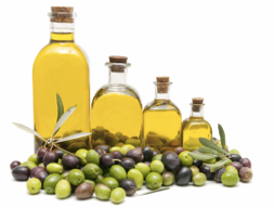
Good sources: Vegetable oils, margarine, wheat germ, nuts, dark green veggies, and whole grains. Liver and eggs contain moderate amounts.
RDA: 10 mg for males and 8 mg for females. However, as your intake of polyunsaturated fatty acids goes up so does the requirement for vitamin E. Remember, the RDA is set to 20% above the level that would cause deficiency symptoms. I think most experts would recommend at least 400 mg/d.
Functions: Vitamin E is an antioxidant (see oxidative damage) that protects cell membranes and other fat-soluble compounds from oxidative damage by free radicals. For example, the oxidative damage to LDL-cholesterol appears to lead to the deposition of cholesterol in the arterial wall leading to atherosclerotic disease. In the past few years many other functions of vitamin E have been clarified. Alpha-tocopherol has direct effect on the control of inflammation, red and white blood cell production, connective tissue growth and genetic control of cell division. Vitamin E acts to reduce free radical damage by converting arachidonic acid is converted to pro-inflammatory (12-HPETE) derivatives. In deficiencies of vitamin E, arachidonic acid is converted to pro-inflammatory leukotrienes and cytokines. In neutralizing free radicals, vitamin E is oxidized to a free radical. Conversion back to the reduced form occurs by reaction with vitamin C (ascorbate).
Deficiency: Almost exclusively limited to premature infants. When it is seen in adults it is usually due to problems with lipid absorption. Symptoms include RBC sensitivity to peroxides and damaged cellular membranes. This is probably due to vitamin E’s antioxidant, protective role.
The principle use of vitamin E is an antioxidant in the protection against heart disease, cancer, stroke and neurodegenerative disease (Alzheimer’s). In addition, alpha-tocopherol supplementation is useful in treating other cardiovascular diseases, diabetes, fibrocystic breast disease, menopause symptoms and tardive dyskinesia. It may also have applications in Parkinson’s Disease and arthritis. Vitamin E is important to immune function, protecting thymic function and white blood cells from oxidative stress.
Symptoms of vitamin E deficiency include nerve damage, muscle weakness, poor coordination, involuntary eye movements, red blood cell fragility, anemia and retrolental fibroplasia (eye disease).
Toxicity: Almost impossible! No toxicity has been noted even at doses 10,000 times the RDA.
Good sources: Green leafy veggies, fruit (esp. strawberries), spinach, cabbage, egg yolks, fish, liver, and dairy products. A large portion of vitamin K is also produced by flora in the intestines. In fact, prolonged treatment with wide-spectrum antibiotics can decrease blood coagulation. Some 2nd generation cephalosporins (cefoperazone, cefamandole, and moxalactam) can also cause this effect (probably due to a warfarin like mechanism).
RDA: No RDA has been set but 70-40 mg per day appears to be adequate.
Functions: The primary function of vitamin K is to aid in the formation of clotting factors and bone proteins. It serves as a cofactor in the production of six proteins that regulate blood clotting, including prothrombin. In addition, it helps to form osteocalcin, a protein necessary for the mineralization of bone. Vitamin K also aids in the formation of glucose into glycogen for storage in the liver. In addition, it promotes the prevention and reversal of arterial calcification, plague progression and lipid peroxidation. Deficiency may increase the risk of calcification of arterial walls, particularly in individuals on vitamin D supplementation (Vitamin D promotes calcium absorption). Vitamin K exists in three forms: K1, a natural form found in plants (phylloquinone); K2, which is synthesized in the intestine (menaquinone); and K3, a synthetic form that must be activated in the liver (menadione). Vitamin K is absorbed in the upper small intestines and transported throughout the body in chylomicrons.
Formation of γ-carboxyglutamate- The proteins for prothrombin and the clotting factors VII, IX, and X are formed as inactive precursors. Carboxylation of glutamate residues forms a mature clotting factor that contains γ-carboxyglutamate (Gla). This carboxylation is dependant upon vitamin K. Dicumarol, a naturally occuring anti-coagulant found in spoilt sweet clover, and warfarin, a synthetic analog of vitamin K, both inhibit the formation of Gla.
Interaction of prothrombin with platelets- The Gla residues contain carboxyl groups (which are negatively charged). These carboxyl groups attract calcium ions (positively charged) which are then able to interact with the phospholipid membranes of platelets.
Role of γ-carboxyglutamate residues in other proteins- These Gla residues are also present in other proteins but the role of vitamin K in their synthesis is not clear.
Deficiency: Hemorrhage. Infants are given an injection of vitamin K at birth. Their intestines are sterile and they cannot have vitamin K produced (hence the injection). However, deficiency is rare in the adult because of the large amount synthesized by intestinal bacteria.
Excessive bleeding, a history of bruising, appearance of ruptured capillaries or menorrhagia (heavy periods) are the most common clinical symptoms of overt vitamin K deficiency, although subclinical deficiency may not affect clotting mechanisms. Due to its critical role in bone formation, long-term vitamin K deficiency may impair bone integrity and growth, eventually predisposing a person to osteoporosis. Antibiotic usage can induce vitamin K deficiency since it upsets the balance of normal intestinal flora. Anticoagulants such as Coumadin and warfarin can also deplete vitamin K by blocking the activation of prothrombin. However, patients on warfarin or other blood anticoagulants should not supplement with vitamin K unless specifically recommended and approved by their physician. Other causes of deficiency include celiac disease, liver disease, certain medications (i.e. aspirin, Dilantin), very high doses of vitamins A and E (over 600 IU) and gastrointestinal disorders associated with the malabsorption of fats, such as bile duct obstruction, pancreatitis or inflammatory bowel disease.
Toxicity: Toxic to the membrane of RBC’s at high doses. Long-term, high-dose administration can cause jaundice and hemolytic anemia.